
![]()
Transmitting Encoded Data Messages from the Field of Master Mind Biology (MMBIO)
Shared from the research of: Joseph Mercado
Content Contributor: Lumen Learning
To: Biology Lover
Blog Post #1182
Re: Molecular Signals and Cellular Receptors
Date and Time: Tuesday, October 5, 2021 at 1:51 a.m.
Dear Biology Lover,
Cellular communication ensures regulation of biological processes within various environments from single-celled to multicellular organisms.
Key Points:
The ability of cells to communicate through chemical signals originated in single cells and was essential for the evolution of multicellular organisms.
In multicellular organisms, cells send and receive chemical messages constantly to coordinate the actions of distant organs, tissues, and cells.
Cells can receive a message, transfer the information across the plasma membrane, and then produce changes within the cell in response to the message.
Single-celled organisms, like yeast and bacteria, communicate with each other to aid in mating and coordination.
Cellular communication has developed as a means to communicate with the environment, produce biological changes, and, if necessary, ensure survival.
Key Terms:
biofilm: a thin film of mucus created by and containing a colony of bacteria and other microorganisms
Introduction: Signaling Molecules and Cellular Receptors
Imagine what life would be like if you and the people around you could not communicate.
You would not be able to express your wishes to others, nor could you ask questions to find out more about your environment.
Social organization is dependent on communication between the individuals that comprise that society; without communication, society would fall apart.
As with people, it is vital for individual cells to be able to interact with their environment. This is true whether a cell is growing by itself in a pond or is one of many cells that form a larger organism.
In order to properly respond to external stimuli, cells have developed complex mechanisms of communication that can receive a message, transfer the information across the plasma membrane, and then produce changes within the cell in response to the message.
In multicellular organisms, cells send and receive chemical messages constantly to coordinate the actions of distant organs, tissues, and cells. The ability to send messages quickly and efficiently enables cells to coordinate and fine-tune their functions.
While the necessity for cellular communication in larger organisms seems obvious, even single-celled organisms communicate with each other. Yeast cells signal each other to aid mating.
Some forms of bacteria coordinate their actions in order to form large complexes called biofilms or to organize the production of toxins to remove competing organisms.
The ability of cells to communicate through chemical signals originated in single cells and was essential for the evolution of multicellular organisms. The efficient and error-free function of communication systems is vital for all forms of life.
Forms of Signaling:
The major types of signaling mechanisms that occur in multicellular organisms are paracrine, endocrine, autocrine, and direct signaling.
Key Points:
Cells communicate via various types of signaling that allow chemicals to travel to target sites in order to elicit a response.
Paracrine signaling occurs between local cells where the signals elicit quick responses and last only a short amount of time due to the degradation of the paracrine ligands.
Endocrine signaling occurs between distant cells and is mediated by hormones released from specific endocrine cells that travel to target cells, producing a slower, long-lasting response.
Autocrine signals are produced by signaling cells that can also bind to the ligand that is released, which means the signaling cell and the target cell can be the same or a similar cell.
Direct signaling can occur by transferring signaling molecules across gap junctions between neighboring cells.
Key Terms:
- Endocrine signaling: signals from distant cells that originate from endocrine cells, usually producing a slow response, but having a long-lasting effect
- Autocrine signaling: produced by signaling cells that can also bind to the ligand that is released: the signaling cell and the target cell can be the same or a similar cell (prefix auto- means self)
- Paracrine signaling: a form of cell signaling in which the target cell is near (para = near) the signal-releasing cell
Forms of Signaling:
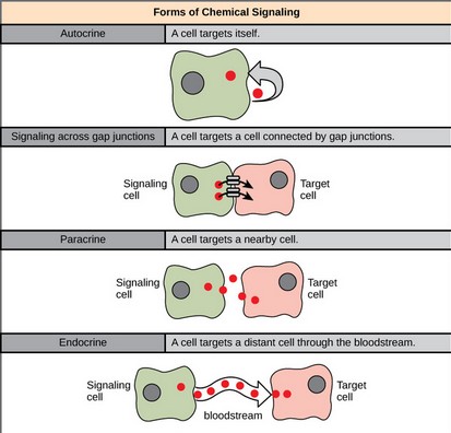
There are four categories of chemical signaling found in multicellular organisms: paracrine signaling, endocrine signaling, autocrine signaling, and direct signaling across gap junctions.
The main difference between the different categories of signaling is the distance that the signal travels through the organism to reach the target cell. It is also important to note that not all cells are affected by the same signals.
image
Forms of Chemical Signaling: In chemical signaling, a cell may target itself (autocrine signaling), a cell connected by gap junctions, a nearby cell (paracrine signaling), or a distant cell (endocrine signaling).
Paracrine signaling acts on nearby cells, endocrine signaling uses the circulatory system to transport ligands, and autocrine signaling acts on the signaling cell. Signaling via gap junctions involves signaling molecules moving directly between adjacent cells.
Paracrine Signaling:
Signals that act locally between cells that are close together are called paracrine signals. Paracrine signals move by diffusion through the extracellular matrix.
These types of signals usually elicit quick responses that last only a short amount of time. In order to keep the response localized, paracrine ligand molecules are normally quickly degraded by enzymes or removed by neighboring cells.
Removing the signals will reestablish the concentration gradient for the signal, allowing them to quickly diffuse through the intracellular space if released again.
One example of paracrine signaling is the transfer of signals across synapses between nerve cells.
A nerve cell consists of a cell body, several short, branched extensions called dendrites that receive stimuli, and a long extension called an axon, which transmits signals to other nerve cells or muscle cells.
The junction between nerve cells where signal transmission occurs is called a synapse. A synaptic signal is a chemical signal that travels between nerve cells. Signals within the nerve cells are propagated by fast-moving electrical impulses.
When these impulses reach the end of the axon, the signal continues on to a dendrite of the next cell by the release of chemical ligands called neurotransmitters by the presynaptic cell (the cell emitting the signal).
The neurotransmitters are transported across the very small distances between nerve cells, which are called chemical synapses. The small distance between nerve cells allows the signal to travel quickly; this enables an immediate response.
image
Synapsis: The distance between the presynaptic cell and the postsynaptic cell—called the synaptic gap—is very small and allows for rapid diffusion of the neurotransmitter. Enzymes in the synapatic cleft degrade some types of neurotransmitters to terminate the signal.
Endocrine Signaling:
Signals from distant cells are called endocrine signals; they originate from endocrine cells. In the body, many endocrine cells are located in endocrine glands, such as the thyroid gland, the hypothalamus, and the pituitary gland.
These types of signals usually produce a slower response, but have a longer-lasting effect.
The ligands released in endocrine signaling are called hormones, signaling molecules that are produced in one part of the body, but affect other body regions some distance away.
Hormones travel the large distances between endocrine cells and their target cells via the bloodstream, which is a relatively slow way to move throughout the body.
Because of their form of transport, hormones get diluted and are present in low concentrations when they act on their target cells. This is different from paracrine signaling in which local concentrations of ligands can be very high.
Autocrine Signaling:
Autocrine signals are produced by signaling cells that can also bind to the ligand that is released. This means the signaling cell and the target cell can be the same or a similar cell (the prefix auto- means self, a reminder that the signaling cell sends a signal to itself).
This type of signaling often occurs during the early development of an organism to ensure that cells develop into the correct tissues and take on the proper function. Autocrine signaling also regulates pain sensation and inflammatory responses.
Further, if a cell is infected with a virus, the cell can signal itself to undergo programmed cell death, killing the virus in the process. In some cases, neighboring cells of the same type are also influenced by the released ligand.
In embryological development, this process of stimulating a group of neighboring cells may help to direct the differentiation of identical cells into the same cell type, thus ensuring the proper developmental outcome.
Direct Signaling Across Gap Junctions:
Gap junctions in animals and plasmodesmata in plants are connections between the plasma membranes of neighboring cells.
These water-filled channels allow small signaling molecules, called intracellular mediators, to diffuse between the two cells.
Small molecules, such as calcium ions (Ca2+), are able to move between cells, but large molecules, like proteins and DNA, cannot fit through the channels. The specificity of the channels ensures that the cells remain independent, but can quickly and easily transmit signals.
The transfer of signaling molecules communicates the current state of the cell that is directly next to the target cell; this allows a group of cells to coordinate their response to a signal that only one of them may have received.
In plants, plasmodesmata are ubiquitous, making the entire plant into a giant communication network.
Types of Receptors:
Receptors, either intracellular or cell-surface, bind to specific ligands, which activate numerous cellular processes.
Key Points:
Intracellular receptors are located in the cytoplasm of the cell and are activated by hydrophobic ligand molecules that can pass through the plasma membrane.
- Cell-surface receptors bind to an external ligand molecule and convert an extracellular signal into an intracellular signal.
- Three general categories of cell-surface receptors include: ion -channel, G- protein, and enzyme -linked protein receptors.
- Ion channel -linked receptors bind a ligand and open a channel through the membrane that allows specific ions to pass through.
- G-protein-linked receptors bind a ligand and activate a membrane protein called a G-protein, which then interacts with either an ion channel or an enzyme in the membrane.
- Enzyme-linked receptors are cell-surface receptors with intracellular domains that are associated with an enzyme.
Key Terms:
- Integral protein: a protein molecule (or assembly of proteins) that is permanently attached to the biological membrane
- Transcription: the synthesis of RNA under the direction of DNA
Types of Receptors:
Receptors are protein molecules in the target cell or on its surface that bind ligands. There are two types of receptors: internal receptors and cell-surface receptors.
Internal Receptors:
Internal receptors, also known as intracellular or cytoplasmic receptors, are found in the cytoplasm of the cell and respond to hydrophobic ligand molecules that are able to travel across the plasma membrane. Once inside the cell, many of these molecules bind to proteins that act as regulators of mRNA synthesis to mediate gene expression.
Gene expression is the cellular process of transforming the information in a cell’s DNA into a sequence of amino acids that ultimately forms a protein. When the ligand binds to the internal receptor, a conformational change exposes a DNA-binding site on the protein.
The ligand-receptor complex moves into the nucleus, binds to specific regulatory regions of the chromosomal DNA, and promotes the initiation of transcription. Internal receptors can directly influence gene expression without having to pass the signal on to other receptors or messengers.
image
Intracellular Receptors: Hydrophobic signaling molecules typically diffuse across the plasma membrane and interact with intracellular receptors in the cytoplasm. Many intracellular receptors are transcription factors that interact with DNA in the nucleus and regulate gene expression.
Cell-Surface Receptors:
Cell-surface receptors, also known as transmembrane receptors, are cell surface, membrane-anchored, or integral proteins that bind to external ligand molecules.
This type of receptor spans the plasma membrane and performs signal transduction, converting an extracellular signal into an intracellular signal. Ligands that interact with cell-surface receptors do not have to enter the cell that they affect.
Cell-surface receptors are also called cell-specific proteins or markers because they are specific to individual cell types.
Each cell-surface receptor has three main components: an external ligand-binding domain (extracellular domain), a hydrophobic membrane-spanning region, and an intracellular domain inside the cell. The size and extent of each of these domains vary widely, depending on the type of receptor.
Cell-surface receptors are involved in most of the signaling in multicellular organisms. There are three general categories of cell-surface receptors: ion channel-linked receptors, G-protein-linked receptors, and enzyme-linked receptors.
Ion Channel-Linked Receptors:
Ion channel-linked receptors bind a ligand and open a channel through the membrane that allows specific ions to pass through. To form a channel, this type of cell-surface receptor has an extensive membrane-spanning region.
In order to interact with the phospholipid fatty acid tails that form the center of the plasma membrane, many of the amino acids in the membrane-spanning region are hydrophobic in nature.
Conversely, the amino acids that line the inside of the channel are hydrophilic to allow for the passage of water or ions.
When a ligand binds to the extracellular region of the channel, there is a conformational change in the protein’s structure that allows ions such as sodium, calcium, magnesium, and hydrogen to pass through.
G-Protein Linked Receptors:
G-protein-linked receptors bind a ligand and activate a membrane protein called a G-protein. The activated G-protein then interacts with either an ion channel or an enzyme in the membrane.
All G-protein-linked receptors have seven transmembrane domains, but each receptor has its own specific extracellular domain and G-protein-binding site.
Cell signaling using G-protein-linked receptors occurs as a cyclic series of events. Before the ligand binds, the inactive G-protein can bind to a newly-revealed site on the receptor specific for its binding.
Once the G-protein binds to the receptor, the resultant shape change activates the G-protein, which releases GDP and picks up GTP.
The subunits of the G-protein then split into the α subunit and the β subunit. One or both of these G-protein fragments may be able to activate other proteins as a result. Later, the GTP on the active α subunit of the G-protein is hydrolyzed to GDP and the β subunit is deactivated. The subunits reassociate to form the inactive G-protein, and the cycle starts over.
Enzyme-Linked Receptors:
Enzyme-linked receptors are cell-surface receptors with intracellular domains that are associated with an enzyme.
In some cases, the intracellular domain of the receptor itself is an enzyme or the enzyme-linked receptor has an intracellular domain that interacts directly with an enzyme.
The enzyme-linked receptors normally have large extracellular and intracellular domains, but the membrane-spanning region consists of a single alpha-helical region of the peptide strand.
When a ligand binds to the extracellular domain, a signal is transferred through the membrane and activates the enzyme, which sets off a chain of events within the cell that eventually leads to a response. An example of this type of enzyme-linked receptor is the tyrosine kinase receptor. The tyrosine kinase receptor transfers phosphate groups to tyrosine molecules.
Signaling molecules bind to the extracellular domain of two nearby tyrosine kinase receptors, which then dimerize. Phosphates are then added to tyrosine residues on the intracellular domain of the receptors and can then transmit the signal to the next messenger within the cytoplasm.
Signaling Molecules:
Signaling molecules are necessary for the coordination of cellular responses by serving as ligands and binding to cell receptors.
Key Points:
- Signaling molecules can range from small proteins to small ions and can be hydrophobic, water-soluble, or even a gas.
- Hydrophobic signaling molecules ( ligands ) can diffuse through the plasma membrane and bind to internal receptors.
- Water-soluble ligands are unable to pass freely through the plasma membrane due to their polarity and must bind to an extracellular domain of a cell -surface receptor.
Other types of ligands can include gases, such as nitric oxide, which can freely diffuse through the plasma membrane and bind to internal receptors.
Key Terms:
- Ligand: an ion, molecule, or functional group that binds to another chemical entity to form a larger complex
hydrophobic: lacking an affinity for water; unable to absorb, or be wetted by water
Signaling Molecules:
Produced by signaling cells and the subsequent binding to receptors in target cells, ligands act as chemical signals that travel to the target cells to coordinate responses.
The types of molecules that serve as ligands are incredibly varied and range from small proteins to small ions like calcium (Ca2+).
Small Hydrophobic Ligands:
Small hydrophobic ligands can directly diffuse through the plasma membrane and interact with internal receptors. Important members of this class of ligands are the steroid hormones.
Steroids are lipids that have a hydrocarbon skeleton with four fused rings; different steroids have different functional groups attached to the carbon skeleton.
Steroid hormones include the female sex hormone, estradiol, which is a type of estrogen; the male sex hormone, testosterone; and cholesterol, which is an important structural component of biological membranes and a precursor of steroid hormones.
Other hydrophobic hormones include thyroid hormones and vitamin D.
In order to be soluble in blood, hydrophobic ligands must bind to carrier proteins while they are being transported through the bloodstream.
Water-Soluble Ligands:
Water-soluble ligands are polar and, therefore, cannot pass through the plasma membrane unaided; sometimes, they are too large to pass through the membrane at all. Instead, most water-soluble ligands bind to the extracellular domain of cell-surface receptors. Cell-surface receptors include: ion-channel, G-protein, and enzyme-linked protein receptors.
The binding of these ligands to these receptors results in a series of cellular changes. These water soluble ligands are quite diverse and include small molecules, peptides, and proteins.
Other Ligands:
Nitric oxide (NO) is a gas that also acts as a ligand. It is able to diffuse directly across the plasma membrane; one of its roles is to interact with receptors in smooth muscle and induce relaxation of the tissue.
NO has a very short half-life; therefore, it only functions over short distances. Nitroglycerin, a treatment for heart disease, acts by triggering the release of NO, which causes blood vessels to dilate (expand), thus restoring blood flow to the heart.
Content Source: Lumen Learning – Boundless Biology
 Email Us a Message
![]()
 Have a question about this article
Please send us a personal message below and we will serve you momentarily. 
We appreciate you visiting the MMU Global Research Directory
For more blog posts, videos, articles, and to generate more knowledge, please feel free and…
 Fly Over to the MMU Facebook Page with Hoot

 Visit the MMA Facebook Group Today
![]()